
Treat Skin Cancer with Green Light

Green Light Inhibits Melanoma Tumor Growth
Summary
Melanin in the outer layers of the skin is energized by green light, even in the absence of ultraviolet.
In small doses, the green light energy will promote proliferation of new, healthy skin cells and inhibit activity in the melanocytes that are the site of melanoma cancer -- even shrinking melanoma tumors.
In larger doses, green light can destroy melanin and melanocytes, therby resolving hyperpigmentation and improving melanona cancer treatment outcomes.
Melanin is the Most Important Chromophore in Skin
There are a myriad of skin disorders, including exczema, psoriasis, acne, rosacea, vitiligo, keratoses, hyperpigmentation, bed sores, and cancer. All of them are modified by the relationship between melanin in the skin and the light environment, but cancer is the most frightening and melanoma is the most deadly. Although it is less common than other skin cancers, melanoma can be more aggressive, grow faster, and metastasize to other parts of the body at a greater rate than basal cell or squamous cell carcinoma. The word melanoma refers to cancer of the melanocytes -- the specialized skin cells near the boundary of the epidermis and dermis that produce the pigment melanin and darkens outer layers of the skin to protect the underlying layers from damaging ultraviolet (UV) rays. The melanocytes synthesize melanin inside tiny structures called melanosomes, then they transfer that melanin to keratinocytes that produce keratin -- the tough, fibrous protein that makes up hair and gives skin its strength, waterproofing, and protective barrier against physical damage, chemicals, and microbes.
Unlike many other forms of cancer that rely primarily on glucose metabolism, melanoma cells exhibit greater metabolic flexibility. They are capable of using glutamine when glucose is scarce, and even exhibit oxidative phosphorylation (OXPHOS) -- the signature metabolic pathway of the mitochondria. That is, the mitochondria inside the cancerous melanocyte are typically dysfunctional, but unlikely to be entirely disabled.
Keeping in mind that melanin is the most important chromophore in the skin, and that melanin converts light into energy that will power skin cells, perhaps it makes sense that the metabolism of cancerous melanocytes will exhibit some unique characteristics that are not shared by other forms of cancer.
Understanding the origins and lifecycle of melonoma requires decoding the metabolism of the melanocytes as it is modified by the light environment.
The consensus view is that melanoma results from damage to the DNA in the nucleus of the cell caused by overexposure to ultraviolet light. However, the evidence is conflicted.
In one Swedish epidemiological study surveying almost 30,000 women, subjects who reported greater sun exposure did experience higher rates of melanoma, which supports the view that excess UV exposure might be the cause of the cancer. However, among the less than 1% of the women who did develop melanoma, all cause mortality was lowest in those women who reported the greatest sun exposure. That is, sunshine increased the rate of melanoma but also reduced the danger, partly because the women with less sun exposure died from other causes, such as cardiovascular disease. The implication is that sunshine promotes overall health from increased Vitamin D synthesis and other benefits that outweigh the risks of UV damage (Lindqvist et al. 2014, 2016, 2022).
What has been overlooked in the current scientific literature is that melanin absorbs more than just UV light. It is so photosensitive that it absorbs across a broad spectrum of wavelengths including green.
The authors of the Swedish studies never asked about exposures to forest light or tree shade, where the light environment is dominated by green and red/near infrared (NIR). Nonetheless, clinical trials have demonstrated the efficacy of these wavelengths to help repair sun-damaged skin.
Why Light and Why Green?
Photodynamic therapy (PDT) is already a clinically approved treatment for treating sun-damaged skin. It works by applying a light-sensitive molecule called a photosensitizer to the skin prior to exposure to a specific wavelength of light that causes the photosensitizers to generate reactive oxygen species (ROS) that lyse cell membranes and destroy the targeted cells. The treatment protocol requires a doctor to apply the photosensitizer cream to the skin, then expose the area to a specific type of light to encourage destruction of the cancer cells in the area that has been sensitized.
When the procedure uses red light, it can be painful to the patient. For this reason, in 1997 a German research team investigated the efficacy of green light and found that compared to red, it was just as effective for healing solar keratoses and less painful (Fritsch et al. 1997). These findings were later corroborated by a Polish research team that achieved complete remission of actinic keratosis using PDT. Their patients treated with green light also reported less pain than those treated with red (Osiecka et al. 2018).
However, melanoma poses a unique biophysical problem that other skin cancers don't -- the tumor itself is often heavily pigmented. Melanin absorbs and scatters light across the visible spectrum, which normally blocks conventional PDT from reaching deeper cancer cells.
It took another 30 years following the German discovery to develop therapies that exploit the photosensitivity of the melanin itself without the necessity of the photosensitizing agent.
Melanin as the Target
A 2022 in vivo study tested this idea, irradiating B16-F10 melanoma cells and mouse tumors with blue (466 nm), green (532 nm), and red (630 nm) light to compare their effects on melanin. The researchers found that excitation of the melanin under light drives photosensitive reactions that generate singlet oxygen and cause oxidative damage. That is, the photosensitive properties of the melanin itself could be used as the mechanism of destroying the cancerous melanocytes without application of a photosensitizing agent (Hausmann et al. 2022).
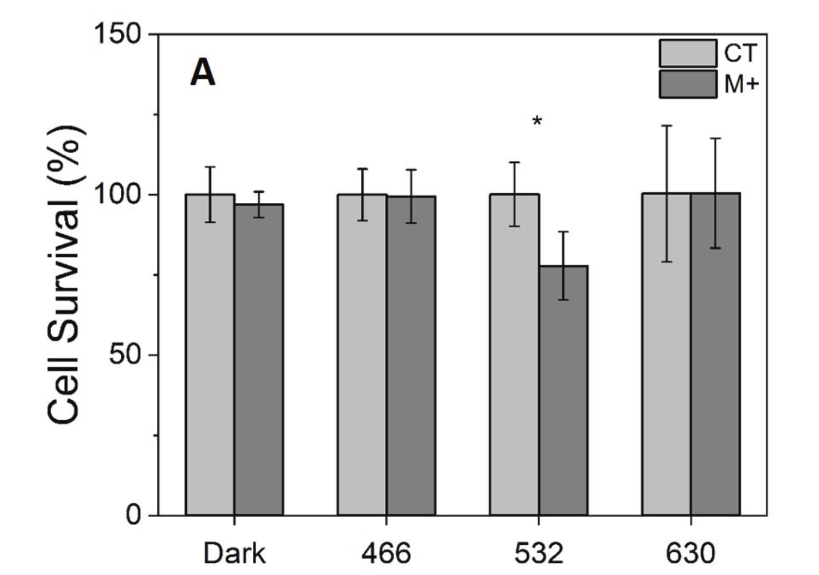
It should be no surprise that among the wavelengths tested, green was the most effective, because melanin is particularly sensitive to green wavelengths of light. As a I wrote in How Green Light Heals Skin, at low levels of exposure the green light will energize melanin, but at higher levels green will destroy it. Moreover, green penetrates skin to the epidermal/dermal boundary at which melanocytes are found.
Green Inhibits Melanocytes
While green light imparts energy to existing melanin that promotes skin healing, it paradoxically inhibits activity in melanocytes. A 2025 Japanese study using 505 nm green LEDs found that irradiation of B16 melanoma cells and 3D skin models reduced melanin production both in cultured cells and in a human trial on facial skin (Yoshihito et al 2025). Moreover, the results were obtained with low-level LED light sources, rather than the higher power and coherent laser light that is typically used to remove age or liver spots (solar lentigines, Zappia et al. 2026).
Because melanoma is an uncontrolled proliferation of cancerous melanocytes, inhibition of their actyivity by green light is a good thing for improving melanoma treatment outcomes, and explain the results of a pioneerring Brazilian study in which mice with induced melanoma were treated with green light and compared to controls that were left untreated. The mice in the experimental group exhibited reduced melanocyte activty, as evidence by both a reduction in tumor volume and a reduction in the concentration of melanin (Haussman et al. 2022).

References
Chen, et al. (2022). The review of the light parameters and mechanisms of Photobiomodulation on melanoma cells. Photodermatology, Photoimmunology & Photomedicine.
Haussmann PB, Pavani C, Marcolongo-Pereira C, Bellettini-Santos T, da Silva BS, Benedito IF, Freitas ML, Baptista MS, Chiarelli-Neto O. Melanin photosensitization by green light reduces melanoma tumor size [mice]. Journal of Photochemistry and Photobiology. 2022 Mar 1;9:100092.
Fritsch C, Stege H, Saalmann G, Goerz G, Ruzicka T, Krutmann J. Green light is effective and less painful than red light in photodynamic therapy of facial solar keratoses. Photodermatology, photoimmunology & photomedicine. 1997 Oct 12;13(5‐6):181-5.
Lindqvist PG, et al. Avoidance of sun exposure is a risk factor for all-cause mortality: results from the Melanoma in Southern Sweden cohort. J Intern Med. 2014;276(1):77–86. doi:10.1111/joim.12251.
Lindqvist PG, et al. Avoidance of sun exposure as a risk factor for major causes of death: a competing risk analysis of the Melanoma in Southern Sweden cohort. J Intern Med. 2016;280(4):375–387. doi:10.1111/joim.12496.
Lindqvist PG, Epstein E, Landin-Olsson M. Sun Exposure – Hazards and Benefits. Anticancer Res. 2022;42(4):1671–1677.
Osiecka BJ, Nockowski P, Szepietowski JC. Treatment of actinic keratosis with photodynamic therapy using red or green light: A comparative study. Acta dermato-venereologica. 2018 Apr 17;98(7):689-93.
Yoshihito MI, Yamada T, Omatsu J, Yamashita T, Suzuki S, Takechi T, Ichikawa M, Yamazaki K, Shinichi SA, Yoshizaki A. Inhibitory effect of 505 nm green light emitting diode on melanin synthesis in cellular experiments and a human intervention study. Acta Dermato-Venereologica. 2025 May 15;105:43441.
Zappia E, Cannarozzo G, Guarino L, Sannino M, Gargano L, Rizzuto G, Clementi A, Duca ED, Dattola A, Pellacani G, Nisticò SP. Use of a 532 nm Green Laser for Solar Lentigines: Case Series and Review. Cosmetics. 2026 May 22;13(3):128.
About the Author
Thomas P Seager, PhD is an Associate Professor in the School of Sustainable Engineering at Arizona State University. He is the founder of MyGreen Lamp, LLC and principal inventor of MyGreen products, and CEO of the Morozko Forge ice bath company.


Comments